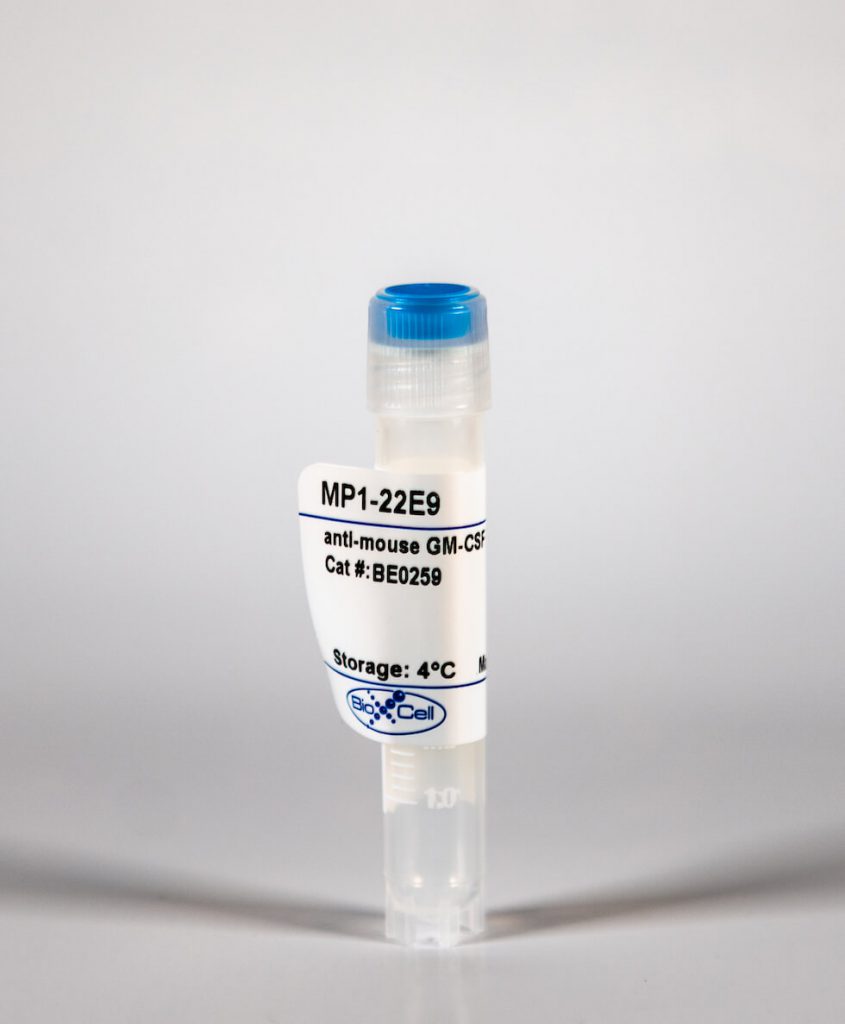
InVivoMab anti-mouse GM-CSF
| Clone | MP1-22E9 | ||||||||||||
|---|---|---|---|---|---|---|---|---|---|---|---|---|---|
| Catalog # | BE0259 | ||||||||||||
| Category | InVivoMab Antibodies | ||||||||||||
| Price |
|
The MP1-22E9 monoclonal antibody reacts with mouse granulocyte-macrophage colony-stimulating factor (GM-CSF), also known as colony stimulating factor 2 (CSF2). GM-CSF is a 14 kDa monomeric hematopoietic factor secreted by macrophages, T cells, mast cells, NK cells, endothelial cells and fibroblasts. GM-CSF stimulates stem cells to differentiate into granulocytes (neutrophils, eosinophils, and basophils) and monocytes. The MP1-22E9 antibody is a GM-CSF neutralizing antibody.
| Isotype | Rat IgG2a, κ |
| Recommended Isotype Control(s) | InVivoMAb rat IgG2a isotype control, anti-trinitrophenol |
| Recommended Dilution Buffer | InVivoPure™ pH 7.0 Dilution Buffer |
| Immunogen | Recombinant mouse GM-CSF |
| Reported Applications |
|
| Formulation |
|
| Endotoxin |
|
| Purity |
|
| Sterility | 0.2 μM filtered |
| Production | Purified from tissue culture supernatant in an animal free facility |
| Purification | Protein G |
| RRID | AB_2687738 |
| Molecular Weight | 150 kDa |
| Storage | The antibody solution should be stored at the stock concentration at 4°C. Do not freeze. |
INVIVOMAB ANTI-MOUSE GM-CSF (CLONE: MP1-22E9)
Kulcsar, K. A., et al. (2014). “Interleukin 10 modulation of pathogenic Th17 cells during fatal alphavirus encephalomyelitis.” Proc Natl Acad Sci U S A 111(45): 16053-16058. PubMed
Mosquito-borne alphaviruses are important causes of epidemic encephalomyelitis. Neuronal cell death during fatal alphavirus encephalomyelitis is immune-mediated; however, the types of cells involved and their regulation have not been determined. We show that the virus-induced inflammatory response was accompanied by production of the regulatory cytokine IL-10, and in the absence of IL-10, paralytic disease occurred earlier and mice died faster. To determine the reason for accelerated disease in the absence of IL-10, immune responses in the CNS of IL-10(-/-) and wild-type (WT) mice were compared. There were no differences in the amounts of brain inflammation or peak virus replication; however, IL-10(-/-) animals had accelerated and increased infiltration of CD4(+)IL-17A(+) and CD4(+)IL-17A(+)IFNgamma(+) cells compared with WT animals. Th17 cells infiltrating the brain demonstrated a pathogenic phenotype with the expression of the transcription factor, Tbet, and the production of granzyme B, IL-22, and GM-CSF, with greater production of GM-CSF in IL-10(-/-) mice. Therefore, in fatal alphavirus encephalomyelitis, pathogenic Th17 cells enter the CNS at the onset of neurologic disease and, in the absence of IL-10, appear earlier, develop into Th1/Th17 cells more often, and have greater production of GM-CSF. This study demonstrates a role for pathogenic Th17 cells in fatal viral encephalitis.
Samavedam, U. K., et al. (2014). “GM-CSF modulates autoantibody production and skin blistering in experimental epidermolysis bullosa acquisita.” J Immunol 192(2): 559-571. PubMed
GM-CSF activates hematopoietic cells and recruits neutrophils and macrophages to sites of inflammation. Inhibition of GM-CSF attenuates disease activity in models of chronic inflammatory disease. Effects of GM-CSF blockade were linked to modulation of the effector phase, whereas effects on early pathogenic events, for example, Ab production, have not been identified. To evaluate yet uncharacterized effects of GM-CSF on early pathogenic events in chronic inflammation, we employed immunization-induced epidermolysis bullosa acquisita (EBA), an autoimmune bullous disease caused by autoantibodies to type VII collagen. Compared to wild-type mice, upon immunization, GM-CSF(-/-) mice produced lower serum autoantibody titers, which were associated with reduced neutrophil numbers in draining lymph nodes. The same effect was observed in neutrophil-depleted wild-type mice. Neutrophil depletion in GM-CSF(-/-) mice led to a stronger inhibition, indicating that GM-CSF and neutrophils have additive functions. To characterize the contribution of GM-CSF specifically in the effector phase of EBA, disease was induced by transfer of anti-type VII collagen IgG into mice. We observed an increased GM-CSF expression, and GM-CSF blockade reduced skin blistering. Additionally, GM-CSF enhanced reactive oxygen species release and neutrophil migration in vitro. In immunization-induced murine EBA, treatment with anti-GM-CSF had a beneficial effect on established disease. We demonstrate that GM-CSF modulates both autoantibody production and skin blistering in a prototypical organ-specific autoimmune disease.
Powell, N. D., et al. (2013). “Social stress up-regulates inflammatory gene expression in the leukocyte transcriptome via beta-adrenergic induction of myelopoiesis.” Proc Natl Acad Sci U S A 110(41): 16574-16579. PubMed
Across a variety of adverse life circumstances, such as social isolation and low socioeconomic status, mammalian immune cells have been found to show a conserved transcriptional response to adversity (CTRA) involving increased expression of proinflammatory genes. The present study examines whether such effects might stem in part from the selective up-regulation of a subpopulation of immature proinflammatory monocytes (Ly-6c(high) in mice, CD16(-) in humans) within the circulating leukocyte pool. Transcriptome representation analyses showed relative expansion of the immature proinflammatory monocyte transcriptome in peripheral blood mononuclear cells from people subject to chronic social stress (low socioeconomic status) and mice subject to repeated social defeat. Cellular dissection of the mouse peripheral blood mononuclear cell transcriptome confirmed these results, and promoter-based bioinformatic analyses indicated increased activity of transcription factors involved in early myeloid lineage differentiation and proinflammatory effector function (PU.1, NF-kappaB, EGR1, MZF1, NRF2). Analysis of bone marrow hematopoiesis confirmed increased myelopoietic output of Ly-6c(high) monocytes and Ly-6c(intermediate) granulocytes in mice subject to repeated social defeat, and these effects were blocked by pharmacologic antagonists of beta-adrenoreceptors and the myelopoietic growth factor GM-CSF. These results suggest that sympathetic nervous system-induced up-regulation of myelopoiesis mediates the proinflammatory component of the leukocyte CTRA dynamic and may contribute to the increased risk of inflammation-related disease associated with adverse social conditions.
Subramanian Vignesh, K., et al. (2013). “Granulocyte macrophage-colony stimulating factor induced Zn sequestration enhances macrophage superoxide and limits intracellular pathogen survival.” Immunity 39(4): 697-710. PubMed
Macrophages possess numerous mechanisms to combat microbial invasion, including sequestration of essential nutrients, like zinc (Zn). The pleiotropic cytokine granulocyte macrophage-colony stimulating factor (GM-CSF) enhances antimicrobial defenses against intracellular pathogens such as Histoplasma capsulatum, but its mode of action remains elusive. We have found that GM-CSF-activated infected macrophages sequestered labile Zn by inducing binding to metallothioneins (MTs) in a STAT3 and STAT5 transcription-factor-dependent manner. GM-CSF upregulated expression of Zn exporters, Slc30a4 and Slc30a7; the metal was shuttled away from phagosomes and into the Golgi apparatus. This distinctive Zn sequestration strategy elevated phagosomal H(+) channel function and triggered reactive oxygen species generation by NADPH oxidase. Consequently, H. capsulatum was selectively deprived of Zn, thereby halting replication and fostering fungal clearance. GM-CSF mediated Zn sequestration via MTs in vitro and in vivo in mice and in human macrophages. These findings illuminate a GM-CSF-induced Zn-sequestration network that drives phagocyte antimicrobial effector function.
Khajah, M., et al. (2011). “Granulocyte-macrophage colony-stimulating factor (GM-CSF): a chemoattractive agent for murine leukocytes in vivo.” J Leukoc Biol 89(6): 945-953. PubMed
GM-CSF is well recognized as a proliferative agent for hematopoietic cells and exerts a priming function on neutrophils. The aim of this study was to determine if GM-CSF has a role as a neutrophil chemoattractant in vivo and if it can contribute to recruitment during intestinal inflammation. Initial studies in vitro, using the under-agarose gel assay, determined that GM-CSF can induce neutrophil migration at a much lower molar concentration than the fMLP-like peptide WKYMVm (33.5-134 nM vs. 1-10 muM). GM-CSF-induced neutrophil migration was ablated (<95%) using neutrophils derived from GMCSFRbeta(-/-) mice and significantly attenuated by 42% in PI3Kgamma(-/-)neutrophils. In vivo, a significant increase in leukocyte recruitment was observed using intravital microscopy 4 h post-GM-CSF (10 mug/kg) injection, which was comparable with leukocyte recruitment induced by KC (40 mug/kg). GM-CSF-induced recruitment was abolished, and KC-induced recruitment was maintained in GMCSFRbeta(-/-) mice. Furthermore, in vivo migration of extravascular leukocytes was observed toward a gel containing GM-CSF in WT but not GMCSFRbeta(-/-) mice. Finally, in a model of intestinal inflammation (TNBS-induced colitis), colonic neutrophil recruitment, assessed using the MPO assay, was attenuated significantly in anti-GM-CSF-treated mice or GMCSFRbeta(-/-) mice. These data demonstrate that GM-CSF is a potent chemoattractant in vitro and can recruit neutrophils from the microvasculature and induce extravascular migration in vivo in a beta subunit-dependent manner. This property of GM-CSF may contribute significantly to recruitment during intestinal inflammation.


